TMR vs. RPNI: What Is the Difference?
Targeted muscle reinnervation (TMR) and regenerative peripheral nerve interface (RPNI) both give a divided nerve a new target after amputation, preventing painful neuromas and enabling prosthetic control. TMR reroutes the nerve into an existing muscle; RPNI wraps it in a small free muscle graft. Dr. Brian Kelley, trained at the University of Michigan where RPNI was pioneered, explains how the two differ, when each is used, and what the published outcomes show.

Introduction
Targeted Muscle Reinnervation (TMR) and Regenerative Peripheral Nerve Interface (RPNI) are the two procedures that have transformed how surgeons manage the divided nerves left behind after amputation. Both address the same fundamental problem — a nerve cut during amputation has no target, forms a disorganized and painful neuroma, and interferes with comfortable prosthetic use. Both solve it by giving the nerve somewhere physiologic to go. They do so through different surgical strategies, and patients researching their options are often confused about how the two differ and which might apply to them.
I trained in plastic surgery and hand and microsurgery at the University of Michigan, where RPNI was developed by Dr. Paul Cederna and the Neuromuscular Laboratory. I had the opportunity to participate in RPNI surgery during that training, in the program where the technique was pioneered, and peripheral nerve surgery has remained a central focus of my practice and my published work since.1 I now practice as a dual board-certified plastic and hand surgeon in Austin, Texas, as an Affiliate Faculty Professor at Dell Medical School at The University of Texas at Austin, and as a partner at the Seton Ascension Institute for Reconstructive Plastic and Hand Surgery.
This post explains what each procedure is, how they differ, what the published literature shows about their outcomes, and how the choice between them — or the decision to use both — is made.
The Problem Both Procedures Solve
A peripheral nerve that is divided needs a target. In normal anatomy, motor nerves end at neuromuscular junctions in muscle and sensory nerves end at receptors in skin. When amputation severs a nerve and removes its target, the proximal nerve stump still attempts to regenerate — that is what nerve tissue does. With no destination, the regenerating axons form a disorganized cluster called a neuroma.
Neuromas produce two clinical problems. The first is pain — a neuroma is hyperexcitable and generates spontaneous and provoked signals that the brain interprets as sharp, electrical, or burning pain, often experienced in the missing limb (phantom limb pain) or in the residual limb (residual limb pain). The second is functional — the neuroma sits where the residual limb meets the prosthetic socket, and the pressure of the socket against it can make wearing a prosthesis intolerable.
Older approaches — burying the nerve in muscle or bone, cutting it and letting it retract, transposing it away from pressure — were palliative and imperfect. They did not address why neuromas form. TMR and RPNI do, and both also generate usable signals for advanced prosthetic control as a second benefit.
What Is Targeted Muscle Reinnervation (TMR)?
TMR redirects a divided nerve into the motor nerve supply of a nearby expendable muscle. The surgeon identifies a small motor nerve branch entering a target muscle, divides it, and coapts (surgically connects) the larger amputated nerve to that smaller motor branch under magnification. The amputated nerve's regenerating axons then grow into the existing motor nerve pathway and reinnervate the target muscle, which becomes their new functional destination.
TMR was originally developed to enable intuitive control of myoelectric prosthetic devices after major upper extremity amputation. The clinical observation that it also reduced neuroma and phantom limb pain emerged from that early experience and has since been confirmed across multiple studies. TMR is now performed both at the time of amputation, to prevent neuroma formation, and as a treatment for established symptomatic neuromas in patients who had amputation without it.
What Is Regenerative Peripheral Nerve Interface (RPNI)?
RPNI takes a different approach. Rather than connecting the divided nerve to an existing muscle's motor nerve, RPNI implants the end of the divided nerve into a small free graft of the patient's own muscle. The graft — typically a few centimeters long — is harvested from a nearby muscle, and the divided nerve end is wrapped within it and secured.
The biology that follows is the key. The free muscle graft initially survives by diffusion, then becomes revascularized from the surrounding tissue. The implanted nerve sprouts axons into the graft and reinnervates it, creating a new, self-contained, organized neuromuscular target. This prevents the disorganized neuroma that would otherwise form and generates a stable electrical signal when the nerve fires — which can be used to control a prosthesis.
RPNI was developed by Dr. Paul Cederna and colleagues at the University of Michigan, originally as a strategy for prosthetic control, with the recognition that it also prevented and treated neuroma pain following from that work. The first clinical series of RPNI for symptomatic postamputation neuromas was reported by the Michigan group, and the technique has since been adopted at peripheral nerve centers internationally.
The Core Difference, Stated Simply
The simplest way to understand the difference: TMR reroutes a nerve into an existing muscle by connecting it to that muscle's nerve supply. RPNI creates a new miniature muscle target by wrapping the nerve in a small free muscle graft.
TMR uses an existing muscle and its existing nerve. RPNI creates a new, self-contained target from transplanted muscle tissue. Both give the divided nerve a physiologic destination, which is what prevents the painful neuroma and generates the usable prosthetic control signal. The strategies differ; the underlying goal is the same.
A few practical distinctions follow from this:
TMR is often selected for major proximal amputations where multiple large nerves need targets and where expendable motor nerve branches in nearby muscles are conveniently available. RPNI is often selected for smaller nerves, for digit-level amputations, for revision of established neuromas, and for situations where a contained, controllable target is preferable. The two are not mutually exclusive — many surgeons use both techniques in the same patient depending on which nerves are being addressed, and combining them is increasingly common.2
When Each Is Performed: Prophylactic vs. Therapeutic
Both procedures can be performed in two settings, and the distinction matters for outcomes.
Prophylactic (also called primary) surgery is performed at the time of amputation, before any neuroma has formed. The divided nerves are managed with TMR or RPNI immediately, preventing the neuroma from developing in the first place. The published literature consistently shows that prophylactic management produces better outcomes than waiting for a neuroma to develop and treating it later.3
Therapeutic (also called secondary) surgery is performed months to years after amputation, in a patient who has developed a symptomatic neuroma or persistent phantom or residual limb pain. The existing neuroma is excised, and the freshened nerve end is then managed with TMR or RPNI. Therapeutic surgery is worthwhile and supported by the literature, though outcomes are generally somewhat less favorable than prophylactic surgery because the nerve has already undergone disorganized regeneration.
What the Literature Shows About Outcomes
The published evidence base for TMR and RPNI has grown substantially and is generally favorable, with some important caveats about study design.
A 2024 systematic review and meta-analysis comparing TMR and RPNI against standard management in limb amputation evaluated the incidence and severity of phantom limb pain, residual limb pain, and painful neuroma, and found that nerve interface procedures performed at the time of amputation reduced these outcomes relative to standard treatment.4 A 2023 systematic review quantifying pain reduction for both prophylactic and therapeutic applications found meaningful reductions in pain across the studies reviewed.5
In the oncologic amputee population specifically — patients who undergo amputation as part of cancer treatment — a 2023 cohort study found that TMR and RPNI were safe and were associated with significant reductions in residual limb pain and phantom limb pain, alongside improvements in patient-reported outcomes. In that study, patient opioid use decreased from approximately 86% preoperatively to approximately 38% postoperatively.6 A separate 2024 case-control study examining combined TMR and RPNI found reductions in long-term narcotic use among amputees who underwent combined nerve management.7
The caveats are honest and important. Much of the published literature consists of cohort studies, case-control studies, and systematic reviews of these — there is limited high-quality randomized data directly comparing TMR to RPNI head-to-head. Follow-up duration varies. Patient populations and outcome measures differ across studies, which complicates direct comparison. The consistent direction of the evidence is favorable, but the field would benefit from more randomized data, and patients should understand that these procedures, while well-adopted, are supported by an evidence base that is still maturing.
Outcomes for Prosthetic Control
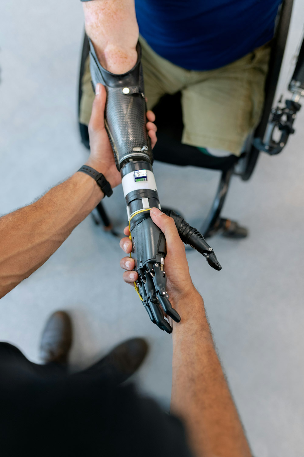
Beyond pain, both procedures generate electrical signals usable for advanced myoelectric prosthetic control. When a patient with a major amputation thinks about moving the missing limb, the brain still sends the motor signal down the relevant nerve. After TMR, that signal reaches the reinnervated muscle; after RPNI, it reaches the implanted muscle graft. The muscle contracts, and surface electrodes in the prosthetic socket detect the resulting electrical signal and translate it into prosthetic movement. The control feels intuitive because the underlying neural pathway is the patient's own. This integration is the foundation of the most advanced upper extremity prosthetic devices in current clinical use.
Risks
Both TMR and RPNI are generally well tolerated, with risk profiles similar to other peripheral nerve operations. Recognized risks include bleeding, infection, persistent or recurrent pain, scarring around the surgical site, incomplete relief of symptoms despite a technically successful operation, and the rare possibility of new pain at the operative site. RPNI involves harvesting a small free muscle graft, which produces a minor and generally inconsequential change at the donor muscle. Both procedures require a recovery period before the biological reorganization is complete and the benefits are fully realized.
Neither procedure is guaranteed to eliminate pain. Some patients have residual pain after TMR or RPNI, and patients with severe long-standing neuromas, multiple prior surgeries, or other chronic pain contributors may experience partial rather than complete relief. Honest counseling about the realistic range of outcomes is part of the decision to operate.
How the Choice Is Made
For a given patient, the choice between TMR, RPNI, or both depends on several factors: the level of amputation, the number and size of nerves to be addressed, the anatomy at the operative site, whether the surgery is prophylactic or therapeutic, and the patient's goals for prosthetic control. There is no universal answer that one technique is superior. The growing practice at many peripheral nerve centers is to use both techniques as the situation calls for them — TMR for the large nerves with convenient motor targets, RPNI for smaller nerves or where a contained target is preferable — and the combination is supported by the outcomes literature.
The decision is best made in consultation with a surgeon experienced in peripheral nerve surgery, ideally as part of a multidisciplinary team that includes the amputation surgeon, prosthetists, and rehabilitation specialists. The timing matters — the strong advantage of prophylactic surgery means that the conversation about nerve management ideally happens before a planned amputation, not after a neuroma has developed.
Related Topics
- Peripheral nerve surgery, RPNI, and TMR
- TMR and RPNI for nerve pain and amputations
- Nerve repair and reconstruction
- Nerve and tendon transfers for nerve injury
- Replantation and revascularization
- RPNI Recovery Timeline and Surgical Guide
- Breast Sensation After Mastectomy
Closing Disclaimer
This article is educational and does not establish a doctor-patient relationship. It does not replace individualized consultation, examination, or review of personal medical history. Patients with amputation-related pain, neuromas, or prosthetic control concerns are encouraged to schedule a consultation with a surgeon experienced in peripheral nerve surgery to discuss their specific situation and options.
Frequently Asked Questions
TMR connects a divided nerve to the motor nerve of an existing nearby muscle, rerouting the nerve's signals into that muscle. RPNI implants the divided nerve into a small free graft of the patient's own muscle, creating a new self-contained target. Both prevent painful neuromas and generate signals for prosthetic control; the surgical strategy differs.
Neither is universally better. The choice depends on the level of amputation, the size and number of nerves being treated, and the clinical goals. TMR is often used for large nerves with convenient muscle targets; RPNI is often used for smaller nerves or where a contained target is preferable. Many surgeons use both techniques in the same patient, and the combination is well supported in the literature.
Both can reduce the incidence and severity of phantom limb pain and residual limb pain, particularly when performed at the time of amputation. The published meta-analyses support this. They do not eliminate pain in every patient — some patients experience partial rather than complete relief, especially when neuromas are long-standing.
Both can be performed prophylactically at the time of amputation to prevent neuroma formation, or therapeutically months to years later to treat an established neuroma. Outcomes are generally better when performed prophylactically, which is why the conversation about nerve management ideally happens before a planned amputation.
Yes. Both generate electrical signals from reinnervated muscle that can be detected by electrodes in a prosthetic socket and translated into prosthetic movement. This is the basis for intuitive control of advanced myoelectric prostheses. The functional benefit depends on the prosthetic device, the level of amputation, and the rehabilitation process.
Yes. Patients who underwent amputation without TMR or RPNI and later developed symptomatic neuromas or chronic pain can be evaluated for therapeutic surgery. The existing neuroma is excised and the nerve is then managed with TMR or RPNI. Outcomes are favorable in selected patients, though generally somewhat less complete than with prophylactic surgery.
No. Both TMR and RPNI are established techniques performed at peripheral nerve centers internationally, with a substantial and growing published evidence base. The evidence consists largely of cohort studies, case-control studies, and systematic reviews rather than large randomized trials, so the field continues to mature, but the procedures are well adopted and supported.
Medical References
1. Hooper RC, Cederna PS, Brown DL, Haase SC, Waljee JF, Egeland BM, Kelley BP, Kung TA. Regenerative Peripheral Nerve Interfaces for the Management of Symptomatic Hand and Digital Neuromas. Plastic and Reconstructive Surgery — Global Open. 2020;8(6):e2792. PMID: 32766027.
2. Targeted Muscle Reinnervation and Regenerative Peripheral Nerve Interface: The Evolving Landscape in the Treatment of Postamputation Pain and Prosthetics. 2026. PMID: 41549863.
3. Mauch JT, Kao DS, Friedly JL, Liu Y. Targeted muscle reinnervation and regenerative peripheral nerve interfaces for pain prophylaxis and treatment: A systematic review. PM&R. 2023;15(11):1457–1465. PMID: 36965013.
4. Yuan M, Gallo M, Gallo L, Huynh MHQ, McRae M, McRae MC, Thoma A, Coroneos CJ, Voineskos SH. Targeted Muscle Reinnervation and Regenerative Peripheral Nerve Interfaces Versus Standard Management in the Treatment of Limb Amputation: A Systematic Review and Meta-Analysis. Plastic Surgery (Oakville). 2024;32(2):253–264. PMID: 38681253.
5. Kubiak CA, Kemp SWP, Cederna PS. Regenerative Peripheral Nerve Interface for Management of Postamputation Neuroma. JAMA Surgery. 2018;153(7):681–682. PMID: 29847613.
6. Outcomes of Targeted Muscle Reinnervation and Regenerative Peripheral Nerve Interfaces for Chronic Pain Control in the Oncologic Amputee Population. 2023. PMID: 37278406.
7. Combined Targeted Muscle Reinnervation With Regenerative Peripheral Nerve Interfaces Decreases Long-Term Narcotic Use in Amputees: A Case Control Study. 2024. PMID: 38527350.
8. Postoperative Imaging Characteristics of Targeted Muscle Reinnervation and Regenerative Peripheral Nerve Interfaces. 2024. PMCID: PMC13002149.



Want to learn more?
Book a consultation